Dental X-Ray in Singapore: When You May Need One
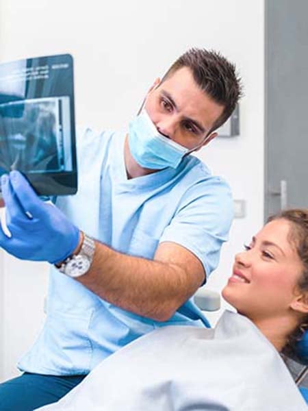
If your dentist has recommended a dental X-ray, you may be wondering whether it is really necessary, how safe it is, or what cost to expect. Some dental problems, including decay between teeth, bone loss, and infections, develop below the gumline in areas that cannot be seen during a standard visual examination. Left undetected, these issues can progress and require more extensive treatment. This guide covers what each type of X-ray involves, when dentists typically recommend them, and what to budget for in Singapore, including available subsidies.
What Are Dental X-Rays?
Dental X-rays, also known as dental radiographs, are diagnostic images that capture detailed views of your teeth, gums, jawbone and surrounding structures. Unlike a standard visual examination, which is limited to what is visible on the surface, dental X-rays allow your dentist to examine areas that cannot be seen with the naked eye, including the spaces between teeth, the roots, the jawbone and the tissues beneath the gumline.
These images are captured using low levels of X-ray radiation, which passes through the soft tissues of the mouth and creates images of the denser structures, such as teeth and bone. They may be part of dental care and are used both for general assessments and for planning specific treatments.
When Does Your Dentist Recommend a Dental X-Ray?
- New patients: At your first visit to the clinic, you may take X-rays to establish a baseline record of your oral health. If you have recent X-rays from another clinic, bring them along, as your dentist may be able to use these instead of taking new ones.
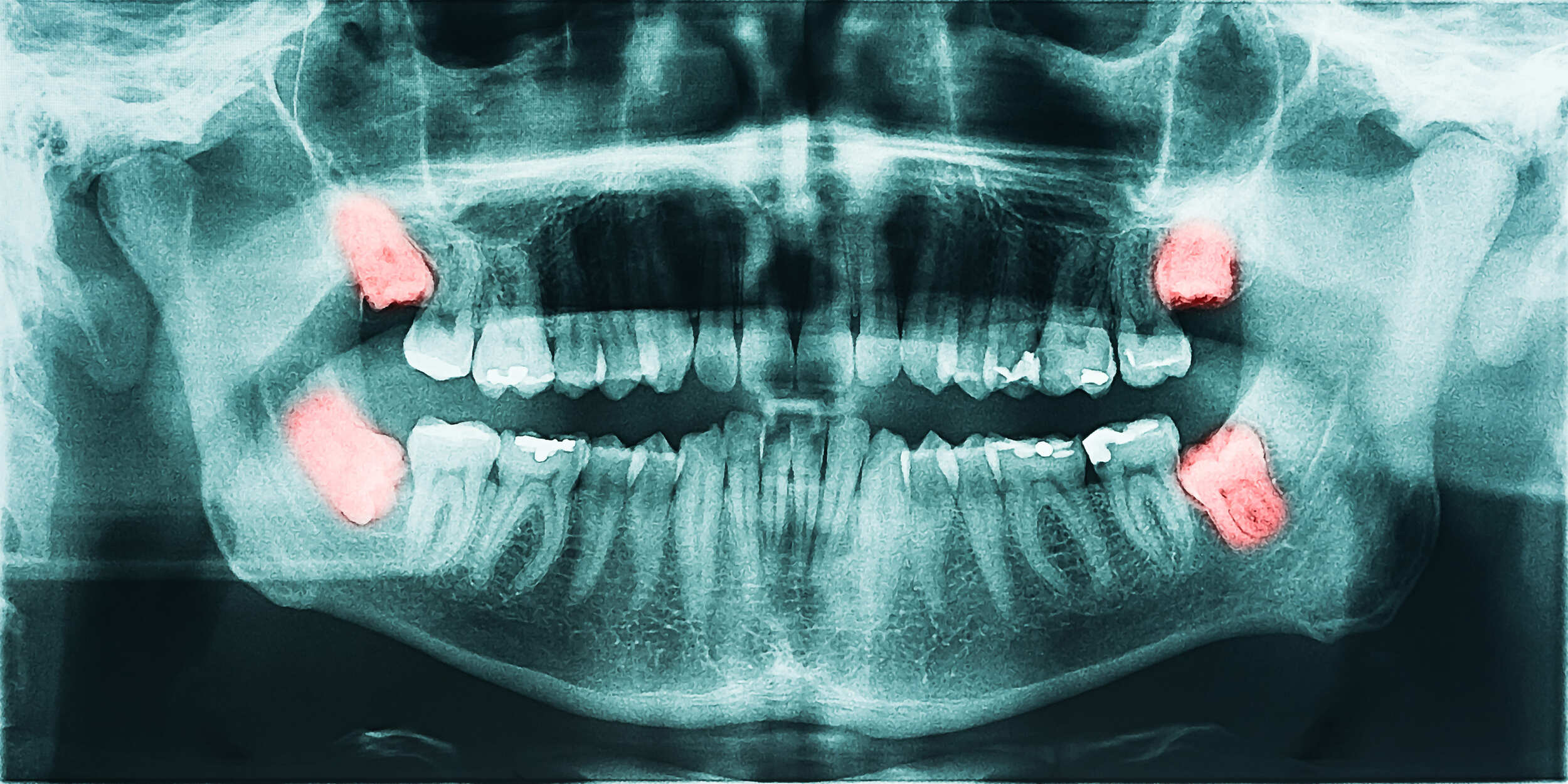
- Pre-treatment assessment: Some procedures require the dentist to assess structures that are not visible during a visual examination, such as bone, tooth roots and nerve position, before treatment begins. X-rays are used to plan procedures including dental implants, wisdom tooth removal, root canal treatment and orthodontics.
- Symptomatic patients: If you are experiencing a toothache, swelling, sensitivity or other oral symptoms, an X-ray helps identify the underlying cause regardless of when you last had one taken.
- Monitoring known conditions: Patients with existing dental issues, such as gum disease or a history of frequent cavities, may require periodic X-rays to track changes and assess treatment response.
- Children's dental development: X-rays help monitor the development of permanent teeth, detect potential crowding, and identify issues such as impacted or congenitally missing teeth early enough for timely intervention.
Dental X-Ray Benefits
Earlier detection of dental issues
X-rays can identify problems such as tooth decay, bone loss and infections before they cause symptoms or become visible during a standard examination. Early detection enables timely intervention, helping accurately identify the root cause and allowing appropriate treatment options to be discussed.
What Can Dental X-Rays Detect?
-
Tooth decay: X-rays can reveal cavities that form between teeth or beneath existing fillings; these are areas that cannot be seen during a visual examination.
-
Root infections and abscesses: Infections at the root of a tooth or in the surrounding bone appear on X-ray images, allowing your dentist to assess the extent of the problem and determine appropriate treatment.
-
Bone loss associated with gum disease: Periodontal disease can cause progressive loss of the bone supporting the teeth. X-rays allow your dentist to assess the level of bone around each tooth and identify changes over time.
-
Impacted teeth: Teeth that have not fully emerged through the gum, including wisdom teeth, can be identified through X-ray imaging. This allows your dentist to assess their position and determine whether intervention is necessary.
-
Cysts, tumours and other abnormalities: Certain cysts, tumours or structural changes within the jawbone or surrounding tissues may be visible on X-ray imaging, depending on the case.
-
Jaw abnormalities and bite problems: Changes to the structure of the jaw, including those associated with temporomandibular joint (TMJ) disorders, can be assessed using specific types of X-rays. X-rays can also reveal problems with how the upper and lower teeth meet, which may affect chewing, jaw function or the fit of dental restorations.
-
Root resorption: During orthodontic treatment, the roots of teeth can shorten over time, a condition known as root resorption. X-rays allow the treating dentist or orthodontist to monitor root length throughout treatment and adjust the treatment plan if necessary.
X-Rays in Treatment Planning
Beyond diagnosis, X-rays are used to plan and guide a range of dental treatments:
-
Dental implants: Bone volume, density and the position of nerves and adjacent teeth are assessed before implant placement.
-
Wisdom tooth removal: The position, angle and proximity of wisdom teeth to surrounding structures such as nerves are evaluated prior to extraction.
-
Root canal treatment: X-rays help map the root canal system and review the treated area after treatment.
-
Orthodontic treatment: The position of teeth, root development and jaw relationships inform treatment planning for braces and aligners.
-
Post-treatment monitoring: Following procedures such as root canal treatment or bone grafting, X-rays allow your dentist to assess healing and monitor changees in the treated area.
Dental X-Ray vs Dental Scan
The term 'dental X-ray' covers all imaging that uses X-ray radiation. Confusion usually arises around two terms: CBCT, which is often called a '3D dental scan', and intraoral digital scanning, which is a different technology altogether.
| Imaging | Uses X-ray radiation? | Notes |
|---|---|---|
| Bitewing, periapical, occlusal, OPG and cephalometric | Yes | The conventional dental X-rays described below |
| CBCT ('3D dental scan') | Yes | Produces 3D images, but is a form of X-ray imaging and involves radiation exposure |
| Intraoral digital scanner | No | Creates digital impressions for orthodontics or restorative work; not an X-ray |
Types of Dental X-Rays
Dental X-rays fall into two main categories: intraoral, where the film or sensor is placed inside the mouth, and extraoral, where imaging is captured outside the mouth. Each type serves a distinct diagnostic purpose.
Intraoral X-Rays
Bitewing X-rays
Bitewing X-rays capture the upper and lower back teeth in a single image, showing the crowns of the teeth and part of the supporting bone. You bite down on a small tab or wing-shaped holder to keep the film in place during the image. These are the most commonly taken X-rays during routine dental visits. They are used primarily to detect decay between teeth, identify decay beneath existing fillings, check the fit of crowns or bridges, and assess early bone changes associated with gum disease.
Periapical X-rays
A periapical X-ray shows the entire length of one or two teeth, from the crown to the tip of the root and the surrounding bone. The sensor or film is placed inside the mouth next to the tooth being examined. These images are used to examine root structure, detect infections or abscesses, identify cysts or other abnormalities at the root tip, and assess the bone supporting the tooth.
Occlusal X-rays
Occlusal X-rays capture the full arch of either the upper or lower teeth in a single image. The film or sensor is held between the biting surfaces of the teeth while the image is taken. They are used to identify extra or missing teeth, locate teeth that have not yet emerged, detect jaw fractures, and identify cysts, abscesses or other pathology. Occlusal X-rays are more commonly used in children to evaluate dental development.
Extraoral X-Rays
Panoramic X-ray (OPG)
A panoramic X-ray, also known as an orthopantomogram (OPG), captures a wide-angle view of the entire mouth in a single image. This includes all the teeth, the upper and lower jaws, the jaw joints and the maxillary sinuses. During the procedure, a rotating arm moves around your head to capture the image. A small bite-blocker is placed between the teeth to ensure proper alignment; no film or sensor is placed inside your mouth.
Panoramic X-rays are used to assess wisdom teeth, evaluate jaw conditions, plan dental implants, detect cysts and tumours, and provide an overview of overall oral health. Because no film is placed inside the mouth, panoramic X-rays may also be recommended for patients with a sensitive gag reflex who find intraoral sensors uncomfortable.
Note on limitations: While the panoramic X-ray provides a broad view, it offers lower resolution than intraoral X-rays such as bitewing or periapical images. For detailed assessment of individual teeth or specific areas of concern, your dentist may recommend supplementary intraoral X-rays alongside the panoramic image.
Lateral Cephalometric X-ray
A lateral cephalometric X-ray captures a side view of the entire head, showing the relationship between the teeth, jaw and facial structure. It is commonly used in orthodontic treatment planning to assess how the teeth and jaws align and to guide decisions about braces or other corrective appliances.
Cone Beam Computed Tomography (CBCT)
CBCT is a form of three-dimensional dental imaging that provides detailed cross-sectional views of the teeth, jaw, nerve pathways, sinuses and surrounding structures (White & Pharoah, 2014). A rotating arm captures data from multiple angles, which is then reconstructed into a 3D image. CBCT is used for complex treatment planning, including dental implants, surgical removal of impacted teeth, assessment of jaw pathology and certain root canal cases. Unlike some forms of CT scanning, CBCT can be performed at a dental clinic rather than a hospital or specialist imaging centre. It delivers more radiation than conventional dental X-rays and is used selectively based on clinical need.
Dental X-Ray Procedure
Before the Procedure
No special preparation is required before a dental X-ray. Your dentist or clinical staff will ask you to:
- Remove any jewellery, glasses or metal objects from the head and neck area, as these can interfere with image quality.
- Inform your dentist if you are pregnant or think you may be pregnant, so appropriate precautions can be taken.
- Let your dentist know of any recent X-rays taken at another clinic.
A lead apron will be placed over your chest and body, and a thyroid collar may be used around your neck to minimise radiation exposure to surrounding tissues.
During the Procedure
The procedure is not expected to be painful and typically takes only a few minutes per image.
For intraoral X-rays, a small sensor or film is positioned inside your mouth next to the teeth being examined. You will be asked to bite down gently or hold the sensor in place while the image is captured. You may feel mild pressure from the sensor, particularly in areas with limited space.
For extraoral X-rays, such as a panoramic or cephalometric X-ray, you will stand or sit still while a rotating arm moves around your head. No film is placed inside your mouth for these types.
It is important to remain as still as possible during the image capture to avoid blurring.
After the Procedure
There is no recovery period following a dental X-ray. You can resume normal activities immediately. Your dentist will review the images, often during the same appointment, and discuss findings and any recommended next steps with you.For digital X-rays, images are usually available shortly after capture and may be reviewed with you during the same appointment.
How Often Should You Get a Dental X-Ray?
There is no single interval that applies to everyone. Your dentist will recommend a frequency based on your oral health history, current condition and risk factors. As a general guide:
Low-risk adults with good oral health and no active dental issues may only need bitewing X-rays (a type of X-ray that shows the crowns of the upper and lower back teeth, covered in detail under Types of Dental X-Rays) every two years, and a full-mouth or panoramic X-ray approximately every three to five years.
High-caries-risk patients: those with a history of frequent tooth decay, active decay or a high bacterial load in the mouth may benefit from more frequent monitoring, typically every six to eighteen months for bitewing X-rays.
Condition-triggered X-rays are taken whenever a new symptom arises, such as a toothache, swelling or sensitivity, without waiting for a scheduled review interval.
Children and adolescents may need X-rays more frequently during periods of active tooth development and growth, particularly where crowding or developmental concerns have been identified.
These intervals are general references and may vary depending on your dentist's clinical assessment.
Dental X-Ray Risks
- Cumulative exposure: Radiation from dental X-rays contributes to lifetime exposure from all sources. Dentists follow the ALARA principle (As Low As Reasonably Achievable), meaning X-rays are only recommended when clinically justified and at the minimum dose necessary for a clear image. In Singapore, X-ray equipment is subject to routine checks and requires an annual licence from the National Environment Agency (NEA).
- Considerations for specific patient groups: Pregnant patients, particularly in the first trimester, are generally advised to postpone routine X-rays unless clinically necessary. When an X-ray is required, a lead apron and thyroid collar are used to reduce exposure to surrounding tissues.
X-rays are recommended based on clinical need and your dentist's assessment.
Dental X-Ray Complications
Discomfort from dental X-rays is uncommon. Possible issues include:
- Gag reflex: Some patients find intraoral sensors uncomfortable, particularly when placed towards the back of the mouth. This may trigger a gag reflex. Informing your dentist in advance allows them to adjust the technique or, where clinically appropriate, recommend a panoramic X-ray instead, which does not require any film placement inside the mouth.
- Discomfort from sensor placement: The edges of an intraoral sensor may cause mild pressure in sensitive areas of the mouth. This is temporary and resolves once the sensor is removed.
Many patients complete dental X-rays with little or no discomfort, although some may experience temporary pressure or gag reflex.
Dental X-Ray Cost in Singapore
The cost of dental X-rays in Singapore varies depending on the type of X-ray, the equipment used and the clinic. The table below provides general market estimates.
| X-Ray Type | Estimated Cost (SGD) |
|---|---|
| Bitewing (per film) | $30 – $60 |
| Periapical (per film) | $30 – $60 |
| Panoramic / OPG | $80 – $150 |
| Lateral Cephalometric | $80 – $150 |
| CBCT (Sectional) | $140 – $220 |
| CBCT (Full) | $300 – $400 |
| Consultation | $27 – $150 |
The figures above are general market estimates in Singapore and are not Nuffield Dental's fee schedule. Actual fees depend on the type of imaging required and the clinic's assessment. The choice of treatment depends on your dental needs, goals, and budget. For more information, please speak with the team at Nuffield Dental.
Please note:
-
All fees are subject to prevailing Singapore Goods and Services Tax.
-
Fees may vary depending on the type and number of images required, the complexity of the procedure, and the type of X-ray or scan recommended.
Are There Subsidies for Dental X-Rays in Singapore?
CHAS
Singaporean Citizens holding a CHAS (Community Health Assist Scheme) card may be eligible for subsidies on dental X-rays at participating CHAS dental clinics. Subsidies are available for up to 6 X-rays per calendar year.
| CHAS Card Type | Subsidy Per X-Ray | Claim Limit |
|---|---|---|
| CHAS Orange | $7.50 | Up to 6 per calendar year |
| CHAS Blue | $11.00 | Up to 6 per calendar year |
| Merdeka Generation (MG) | $16.00 | Up to 6 per calendar year |
| Pioneer Generation (PG) | $21.00 | Up to 6 per calendar year |
To claim CHAS subsidies, treatment must be received at a CHAS-participating dental clinic. Bring your CHAS card to your appointment and check with the clinic on eligibility before proceeding. Information is accurate as at June 2026, and may be updated by CHAS. Please check the CHAS website or confirm with the clinic before your appointment.
MediSave
MediSave generally does not cover dental X-rays, as they are classified as non-surgical dental procedures. MediSave claims for dental treatment are limited to qualifying surgical procedures. For more information on MediSave eligibility, check with your dental clinic or refer to the CPF Board guidelines.
Dental Insurance
Coverage for dental X-rays under private dental insurance varies by policy. Some plans include X-rays as part of a basic dental benefit, while others may apply annual limits or require that X-rays form part of a broader covered procedure. Review your policy documents or contact your insurer to confirm what is covered under your plan.
Why Choose Nuffield Dental for Dental X-Rays?
- Digital imaging technology: Nuffield Dental uses digital X-ray systems across its clinics. Images are produced within seconds, can be adjusted for contrast and brightness, stored for comparison at future visits, and transferred electronically to other providers or specialists as needed. For cases requiring three-dimensional assessment, selected Nuffield Dental clinics may provide CBCT imaging where clinically indicated, which supports planning for procedures such as dental implants, wisdom tooth removal, and root canal treatment. Please check with the clinic for availability.
- Multiple clinic locations across Singapore: Nuffield Dental currently has 10 clinic locations across Singapore. Whether near your home, workplace or school, a clinic is likely within reach. Please refer to the clinic locations page for the current list of locations.
Digital vs Traditional X-Ray
| Digital X-Rays | Traditional (Film) X-Rays | |
|---|---|---|
| Radiation exposure | Generally lower radiation exposure than traditional film | Higher radiation dose required to expose film |
| Image availability | Images can usually be viewed shortly after capture, allowing same-visit review | Film must be chemically developed before viewing |
| Image adjustment | Can be enlarged, and adjusted for contrast and brightness to support diagnosis | Fixed once developed; cannot be adjusted |
| Storage | Stored digitally; easily retrieved for future comparison | Physical film; requires physical storage and can degrade |
Nuffield Dental uses digital X-ray systems at its clinics. Check with your preferred clinic for the imaging services available.
FAQ
Is a dental X-ray painful?
Dental X-rays are typically well tolerated by patients. The procedure involves positioning a small sensor or film holder inside the mouth, which may cause temporary discomfort for some individuals. The X-ray exposure itself takes only a few seconds.
Do I need a dental X-ray at every dental check-up?
No, a dental X-ray is not required at every check-up. Your dentist will assess whether one is needed based on your oral health history, risk factors, and any symptoms. Patients with good oral health and a low risk of dental disease may only need X-rays every one to two years. Those with a higher risk profile or active dental concerns may need them more frequently. Your dentist will advise on the appropriate interval for your individual situation.
Are dental X-rays safe for children?
Dental X-rays are generally considered safe for children when clinically indicated. The radiation dose involved is low, and dentists take care to limit X-rays in younger patients to those that are clinically necessary. The benefit of detecting developmental issues or decay early is weighed against the principle of keeping radiation exposure as low as reasonably achievable.
When is a dental X-ray not recommended?
Your dentist may advise against or postpone a dental X-ray in certain situations, including during the first trimester of pregnancy unless necessary, shortly after a recent set of X-rays where no clinical changes have occurred, or in patients at low risk of dental disease with no symptoms requiring further investigation. Your dentist will weigh the clinical benefit of the X-ray against the level of exposure for your individual situation.
Can I refuse a dental X-ray?
Yes. You have the right to make informed decisions about your dental care, including whether to proceed with an X-ray. However, your dentist may advise that proceeding with certain treatments without X-ray imaging may not be clinically appropriate, as accurate diagnosis depends on the information these images provide. Discuss any concerns with your dentist so that an informed decision can be made together.
Can dental X-rays detect oral cancer?
Dental X-rays may show certain abnormalities in the jawbone, but they do not detect all forms of oral cancer. However, X-rays do not detect all forms of oral cancer, particularly those affecting soft tissues. Regular oral cancer screenings conducted by your dentist, which involve a visual and physical examination of the oral tissues, remain an important part of ongoing oral health monitoring.
How much radiation is there in a dental X-ray?
Dental X-rays involve low levels of radiation, measured in microsieverts (μSv). Typical exposures vary by type:
- Bitewing (4 films): approximately 5 μSv
- Periapical (2 to 4 films): approximately 3 μSv
- Panoramic: approximately 10 to 30 μSv
- Cone Beam CT (CBCT): approximately 100 to 200 μSv
For context, natural background radiation from sources such as sunlight, soil, and air travel amounts to around 3,500 μSv per year. Your dentist will only recommend X-rays when clinically indicated.
The Nuffield Dental Clinic Network In Singapore
Nuffield Dental Seletar
Greenwich V
1 Seletar Road #01-07/08
Singapore 807011
Nuffield Dental Kovan
Simon Plaza
2 Kovan Road #01-03
Singapore 548008
Nuffield Dental Serangoon Gardens
Serangoon Garden Estate
57 Serangoon Garden Way
Singapore 555953
Nuffield Dental Siglap
The Domain
914 East Coast Road #01-03
Singapore 459108
Nuffield Dental Bedok
East Village Mall
430 Upper Changi Road #01-64
Singapore 487048
Nuffield Dental Holland Village
7 Holland Village Way #03-16
Singapore 275748
Nuffield Dental Jurong East
Nuffield Dental Westgate
Westgate
3 Gateway Dr #04-32
Singapore 608532
Nuffield Dental Marina One
The Heart, Marina One
5 Straits View #B2-58
Singapore 018935
Nuffield Dental Orchard
Nuffield Dental Jewel
Wheelock Place
501 Orchard Road #05-01
Singapore 238880
Nuffield Dental Raffles Place
One Raffles Place
1 Raffles Place #05-19
Singapore 048616
Book An Appointment
We would be happy to assist with your enquiry.
Please fill out this form if you would like to book an appointment with us or if you have a question for us. Our team will get in touch after reviewing your submission.
Dental Care at Nuffield Dental
Nuffield Dental is a dental clinic providing general and specialist dental services. We provide dental care based on each patient's individual clinical needs. Our team provides information and support before, during and after treatment.
Nuffield Dental provides dental X-ray imaging as part of dental assessment and treatment planning, where clinically appropriate.
Dental X-rays may be recommended by the attending dental practitioner to assess teeth, gums, jawbone, or other oral structures.
The type of X-ray required will depend on the patient’s clinical condition and the dentist’s assessment.
Articles
The newest and best lifestyle articles selected by our editorial team.
- 11 Feb 2026
- 2 mins read
- Dental Fillings
Why the Global Phase-Down Is Happening Mercury is listed by the WHO as one of the top ten chemicals of public health concern. In dentistry, the...
- 17 Nov 2025
- 2 mins read
Current scientific evidence does not show a conclusive connection between intact dental amalgam fillings and symptoms such as brain fog, chronic...
- 17 Nov 2025
- 2 mins read
The Minamata Convention on Mercury established 2034 as the global target year to end the use of dental amalgam. While this may seem gradual, the...
.png?width=2223&height=447&name=Background%20(4).png)

